Shared validation data

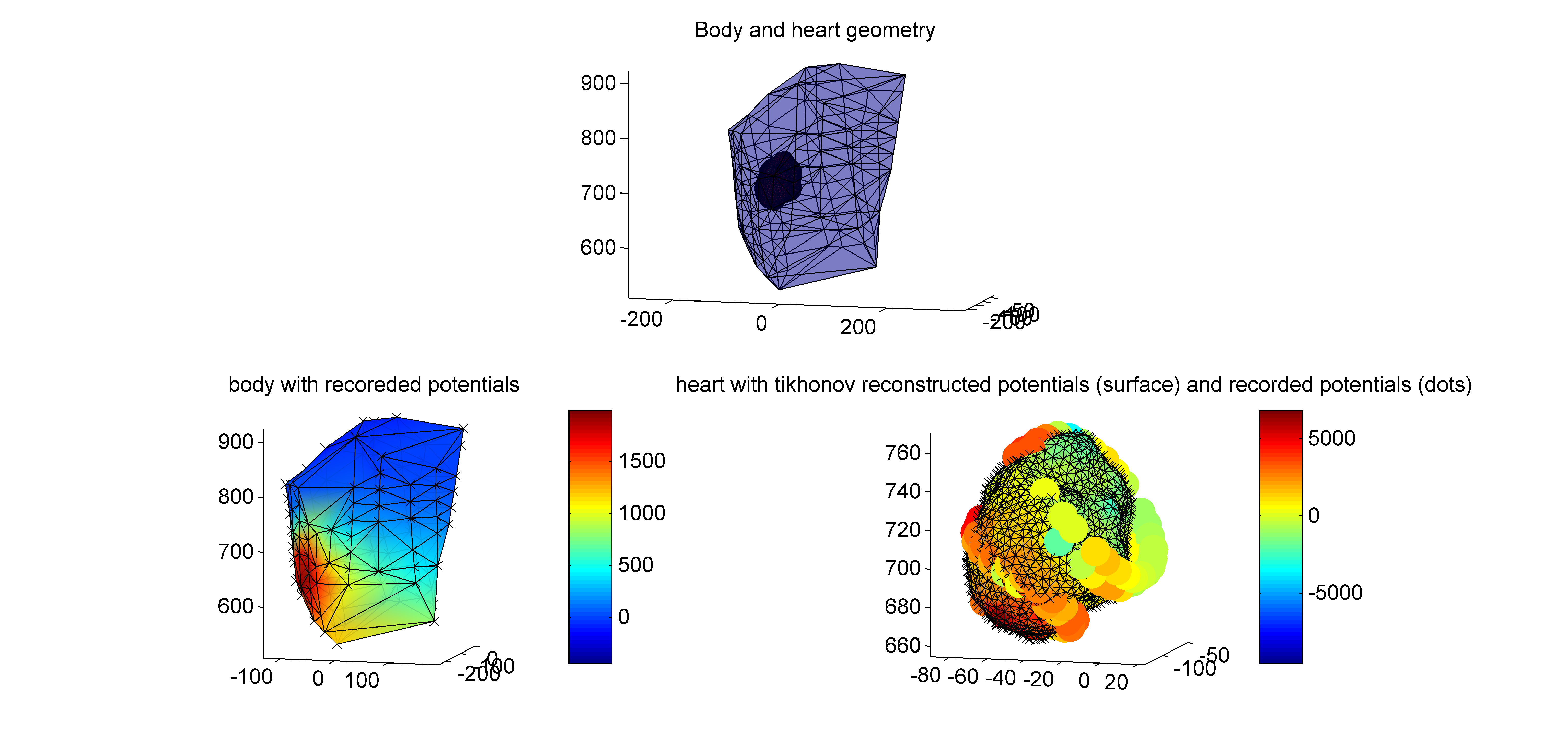
We have been performing validation experiments to validate noninvasive reconstruction of electrocardiographic imaging. In these experiments, we have acquired extensive, simultaneous, in vivo, recordings of body-surface potentials and epicardial potentials. We freely share two data sets, containing the full body-surface geometry (consisting of approx. 140 well-connected body-surface electrodes), the epicardial ventricular surface, and epicardial electrodes (approx. 70 well-connected electrodes). The first data set contains the recording of a sinus beat; the second data set contains recordings of a beat paced epicardially at the left ventricular apex. Some Matlab code provides a demo to illustrate the data. We also provide a software network in SCIrun that allows you to visualize and reconstruct epicardial potentials, based on these data.

To acquire these data sets, please contact me at: m.cluitmans (at) maastrichtuniversity.nl and you’ll receive them by e-mail. Please state your name, affiliation and purpose, and we encourage you to keep us updated on your research with these data. You can also download a subset of these data from the EDGAR repository here.

If you use these data in publications, we require you to cite the following publication:

M. J. M. Cluitmans et al., In Vivo Validation of Electrocardiographic Imaging. JACC: Clinical Electrophysiology, 232–242 (2017).

We hope these data will be fruitful for your research!

The shared data visualized in Matlab with the provided Matlab code. The geometry of heart, body and epicardial electrodes is provided, together with potential recordings.
Example of shared data, consisting of the body-surface electrodes (depicted as a mesh in this image), heart-surface (drawn as a full surface), and epicardial electrodes (spheres). The body-surface potentials and epicardial electrode potentials are recorded and plotted as colors on the respective spheres. The colors of the heart-surface are an example of Tikhonov-reconstructed potentials, that can be compared to the recorded epicardial potentials.
This SCIrun network solves the inverse problem of electrocardiography, based on measured body-surface potentials and the provided geometry.